

Photo courtesy of FUJIFILM Sonosite.
February 12, 2025
Four new ultrasound machines that practitioners are calling “state-of-the-art technology” and “top-notch” are making a difference in cardiac care at St. Boniface Hospital, thanks to the generosity of Foundation donors.
These are in addition to six similar, but older, ultrasounds that were already at St. B. The new Sonosite PX ultrasounds were funded in part through the Foundation’s successful Voices of Hope campaign. Our campaign matching donors tripled gifts made to patient care and medical research at St. B over the last six weeks of 2024. Donors reached the fundraising goal of $250,000 for the four machines, which were delivered to the Hospital in December of 2024.
Clinicians use the ultrasounds in the Section of Cardiac Anesthesia, which deploys theirs in three cardiac operating rooms; for inpatients and outpatients in the Bergen Cardiac Care Centre on the Hospital’s second floor; and in the Cardiology Inpatient and Acute Cardiac Care Units on the fifth floor.
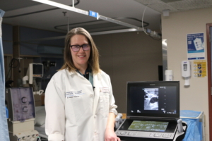
“Now we aren’t taking a resource away from other operating rooms,” said Dr. Mullein Thorleifson, Medical Director of Cardiac Anesthesia at the Hospital. “If we were using ultrasound in cardiac before, it meant that somebody else sharing that machine didn’t have it available for another sick patient,” she said.
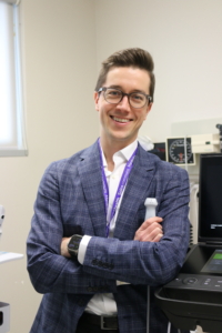
Also called a point-of-care ultrasound system, the portable machines offer fantastic image quality for clinicians, said Dr. Justin Cloutier, Medical Director of the Cardiology Clinical Teaching Units at the Hospital. Having one on each cardiac unit and in the clinic area, the ultrasound is always just a few steps away, he added.
“The resolution of the new ultrasound probes can make the difference between the picture looking like a January blizzard in Winnipeg, and a crisp image where you can confidently make a diagnosis,” said Cloutier.
“We have patients coming in from all over the province with cardiac issues,” he continued. “The ultrasound is a really a window inside to the heart. We can assess ventricular function, look at the valves of the heart, see if there’s fluid accumulating around the heart, and do heart volume status assessments. As a supplement to overall history and physical exam, it’s really a cornerstone of how we do cardiac assessments,” he said.
“It’s all about the safety and comfort of our patients.”
The Sonosite ultrasounds are crucial tools, agreed Dr. Thorleifson. “Before patients come into a cardiac operating room, sometimes we need to use the ultrasound’s cardiac probe to assess their heart function, to make sure we can get people off to sleep safely,” she said.
“We can look at their heart and see what the heart function is like, and it allows us to tailor our anesthetic to that patient.”
Thorleifson uses her unit’s ultrasound “in every single case” to put IVs in patients before and during cardiac surgeries. These include pre-op intravenous and arterial lines, where “using it means that our first pass success is higher, so we’re less likely to poke people multiple times,” she explained.
“It’s all about the safety and comfort of our patients.”
She noted that many cardiac patients have been “poked literally hundreds of times for various procedures,” making it more difficult to put in another IV for the surgery. Patients have to be poked again before they go off to sleep.
The ultrasound gives Thorleifson a quick, clear look at veins deep inside a patient’s inner forearm, a better location where she can put in an IV more comfortably – as opposed to painful spots like the back of a hand or inner elbow.
After the patient is asleep, Thorleifson uses the ultrasound to put in a central line in the neck, and in fascial blocks before the patient wakes up, in which she puts local anesthetic on either side of the chest wall to help manage pain after surgery.
Ultrasounds reducing wait times
The next step up in diagnostic imaging from the Sonosite ultrasounds is a full echocardiogram, explained Dr. Cloutier. “As you can imagine, there are frequently dozens of patients at the Hospital waiting for an ‘echo’,” he pointed out.
The new ultrasounds allow clinicians to make assessments and make plans “right then and there,” he said. “They help us make more quick decisions in care management plans for patients. On the patient side, you’re not sitting necessarily in hospital waiting days for an echocardiogram.”
“After someone’s had a heart attack, there are certain kinds where we may not need that full echocardiogram. But we want to know what their heart function looks like, and the Sonosite ultrasounds facilitate that.”
“Rather than waiting for the echo, I can look at their heart function and give them a verbal diagnosis of whether there’s been extensive heart damage or not. That could mean the patient goes home a day or two earlier,” he said.